Beta, Alpha, Gamma Hemolysis: Understanding Bacterial Growth Patterns

<!DOCTYPE html>
Understanding hemolysis patterns is crucial in microbiology, especially when identifying and classifying bacteria. These patterns—beta, alpha, and gamma hemolysis—provide valuable insights into how bacteria interact with blood cells, aiding in diagnosis and treatment. Whether you’re a student, researcher, or medical professional, grasping these concepts is essential for interpreting lab results accurately.
What is Hemolysis?

Hemolysis refers to the breakdown of red blood cells (RBCs). In microbiology, it’s observed when bacteria are cultured on blood agar plates. The type of hemolysis—beta, alpha, or gamma—depends on how bacteria affect the RBCs. This phenomenon is a key characteristic used to identify bacterial species.
Beta Hemolysis: Complete Breakdown

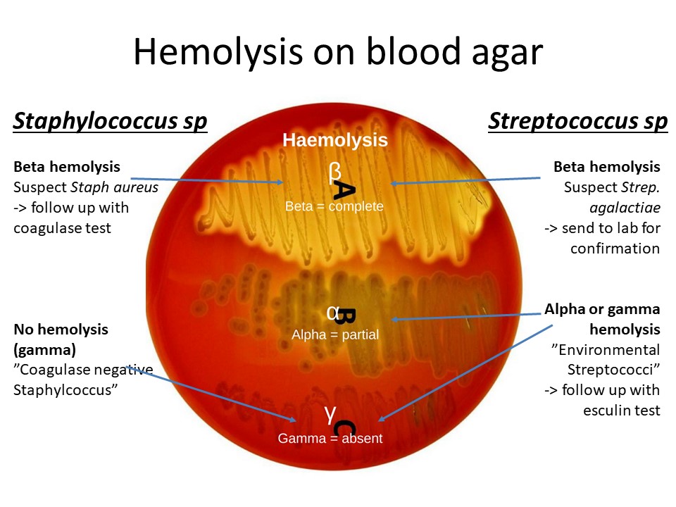
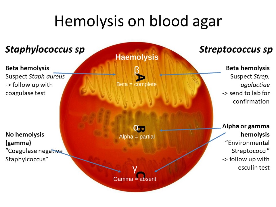
Beta hemolysis is characterized by a clear zone around bacterial colonies on a blood agar plate. This occurs when bacteria produce enzymes that completely lyse RBCs, releasing hemoglobin. Common examples include Streptococcus pyogenes, a pathogen associated with strep throat.
💡 Note: Beta-hemolytic bacteria are often pathogenic and require careful identification and treatment.
Alpha Hemolysis: Partial Breakdown

Alpha hemolysis appears as a green or brown discoloration around the colony, indicating partial lysis of RBCs. This is due to the production of hydrogen peroxide by bacteria like Streptococcus pneumoniae. The oxidation of hemoglobin causes the color change.
Gamma Hemolysis: No Breakdown

Gamma hemolysis shows no visible changes around the bacterial colony. The RBCs remain intact, as seen with bacteria like Staphylococcus epidermidis. This type is often associated with non-pathogenic or less virulent strains.
| Hemolysis Type | Appearance | Example Bacteria |
|---|---|---|
| Beta | Clear zone | Streptococcus pyogenes |
| Alpha | Green/Brown zone | Streptococcus pneumoniae |
| Gamma | No change | Staphylococcus epidermidis |

Why Hemolysis Matters in Microbiology

Hemolysis patterns are vital for:
- Identifying bacterial species
- Understanding bacterial virulence
- Guiding treatment decisions
Checklist for Identifying Hemolysis Patterns
- Observe the blood agar plate for clear, green/brown, or no zones.
- Note the colony appearance and color changes.
- Compare findings with known bacterial characteristics.
Mastering beta, alpha, and gamma hemolysis is fundamental in microbiology, enabling accurate bacterial identification and informed medical decisions. (bacterial identification, blood agar plates, microbiology techniques)
What causes beta hemolysis?
+Beta hemolysis is caused by bacterial enzymes that completely lyse red blood cells, creating a clear zone around the colony.
How is alpha hemolysis different from beta hemolysis?
+Alpha hemolysis results in partial lysis of RBCs, causing a green or brown discoloration, while beta hemolysis creates a clear zone.
Why is gamma hemolysis important?
+Gamma hemolysis indicates no RBC lysis, helping identify non-pathogenic or less virulent bacteria.


