Exploring E. Coli Bacteria Under the Microscope

Exploring the microscopic world of E. coli bacteria can be both fascinating and educational. Whether you're a student, researcher, or simply curious about microbiology, understanding how to observe E. coli under a microscope is a valuable skill. This bacterium, scientifically known as *Escherichia coli*, is commonly found in the intestines of humans and animals. While some strains are harmless, others can cause infections, making it a crucial subject in both medical research and educational studies. In this post, we’ll guide you through the process of preparing and viewing E. coli samples, ensuring you gain a clear and detailed perspective of this microscopic organism.
What You Need to Observe E. Coli Under a Microscope

Essential Equipment for Microscopic Analysis
To begin your exploration of E. coli bacteria, you’ll need a few essential tools. These include a compound microscope, which allows for higher magnification compared to a stereo microscope. Additionally, you’ll require microscope slides, cover slips, and staining materials like Gram stain for better visibility. A Bunsen burner or alcohol lamp is also necessary for sterilizing equipment and fixing the sample. Lastly, ensure you have a culture of E. coli, which can be obtained from a laboratory supplier or prepared using nutrient agar.
Safety Precautions When Handling E. Coli
Working with E. coli requires strict adherence to safety protocols. Always wear protective gloves and a lab coat to prevent contamination. Ensure your workspace is clean and well-ventilated. Avoid touching your face or eating while handling the bacteria. Properly dispose of all materials, including used slides and culture plates, in designated biohazard containers. Following these precautions minimizes the risk of infection and ensures a safe experimental environment.
Step-by-Step Guide to Preparing E. Coli Samples

Culturing E. Coli for Microscopic Observation
Start by preparing a nutrient agar plate to culture E. coli. Sterilize the plate using an autoclave or microwave, then allow it to cool. Inoculate the plate with a small amount of E. coli culture using a sterile loop. Incubate the plate at 37°C for 24 hours to promote bacterial growth. Once colonies are visible, you can proceed to prepare a wet mount for microscopic examination.
Creating a Wet Mount for Microscopy
To create a wet mount, place a drop of sterile water or saline solution on a clean microscope slide. Using a sterile loop, gently scrape a small portion of the E. coli colony from the agar plate and mix it into the drop. Carefully place a cover slip over the sample, ensuring no air bubbles are trapped. This preparation is now ready for observation under the microscope.
📌 Note: Always label your slides and culture plates to avoid confusion and ensure proper tracking of samples.
Observing E. Coli Under the Microscope

Adjusting Microscope Settings for Optimal Viewing
Begin by placing the prepared slide on the microscope stage. Start with the lowest magnification (e.g., 4x or 10x) to locate the bacteria. Gradually increase the magnification to 40x or 100x for a detailed view. Adjust the focus knobs to sharpen the image. If using a compound microscope, ensure the condenser and diaphragm are properly aligned for optimal lighting.
Identifying E. Coli Characteristics
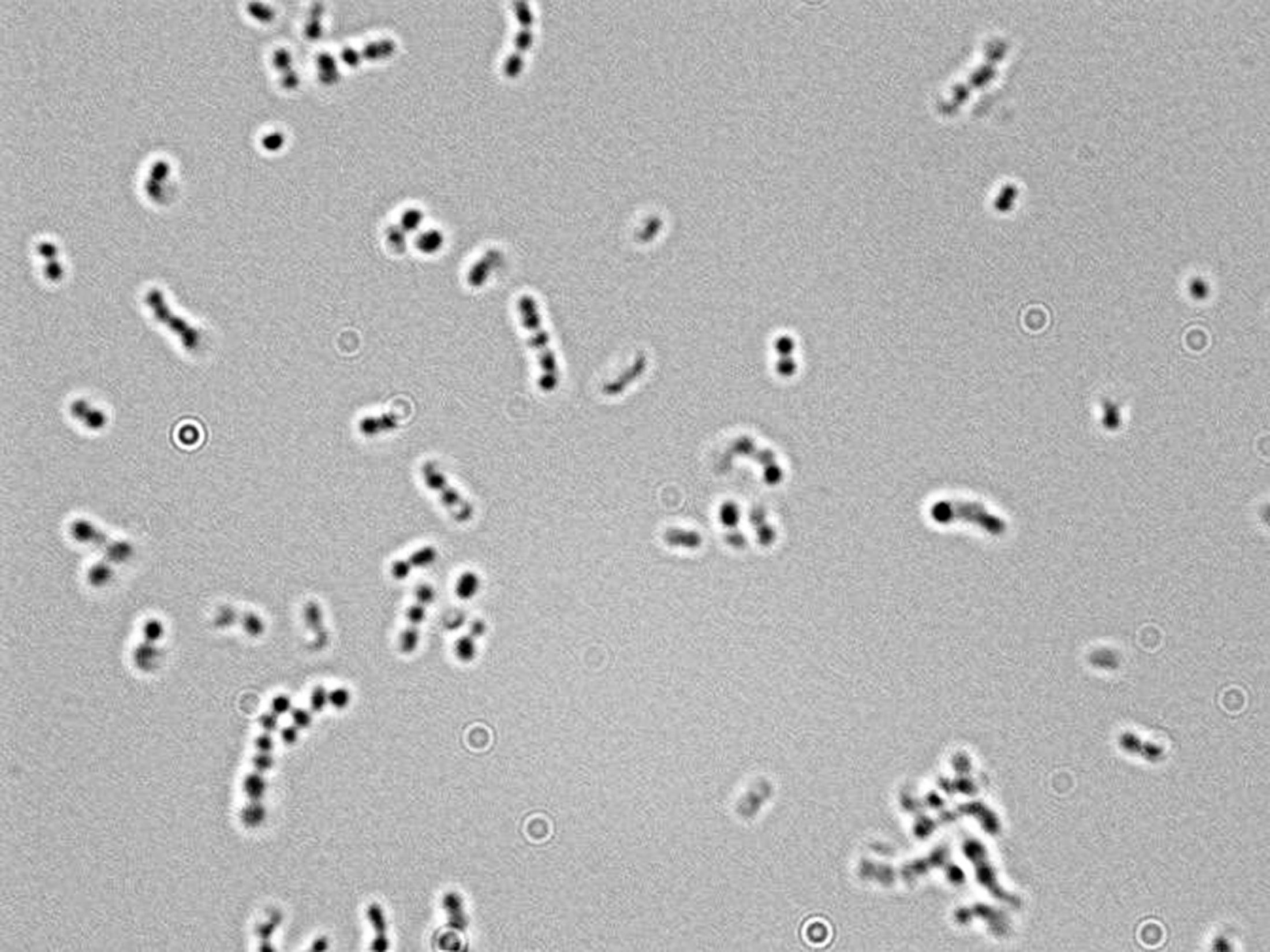
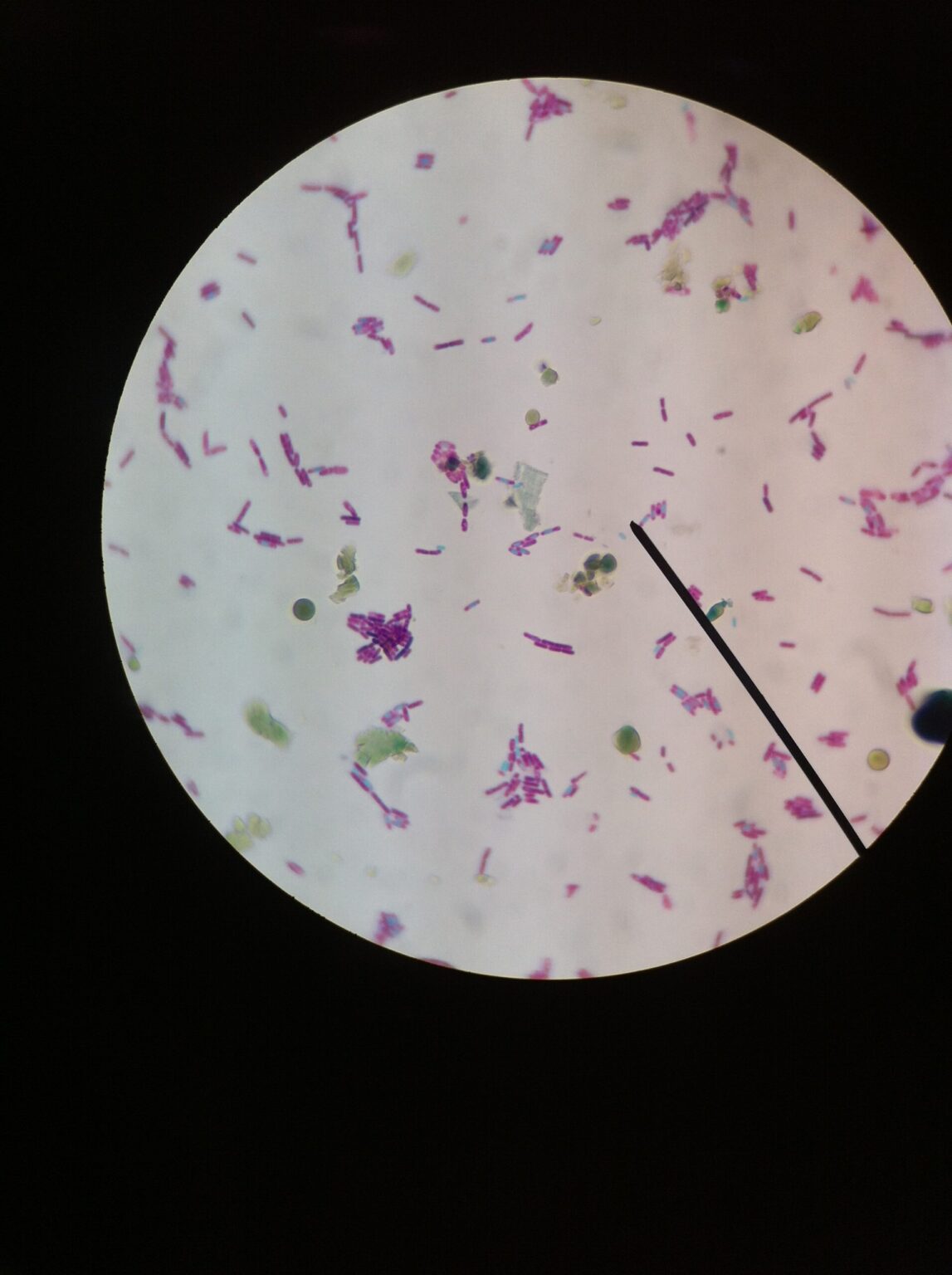
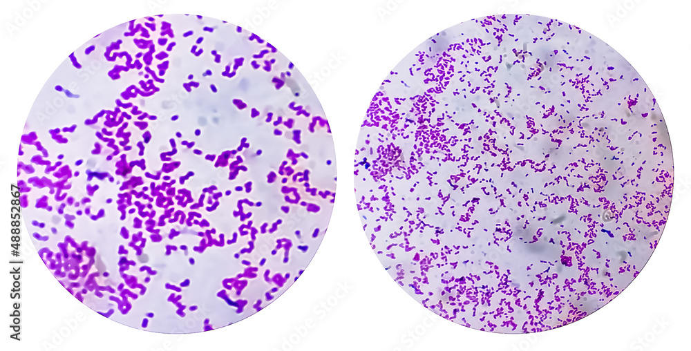
Under the microscope, E. coli appears as small, rod-shaped bacteria, typically measuring around 1-2 micrometers in length. They often occur singly or in pairs. For enhanced visibility, apply a Gram stain, which will color the bacteria pink (Gram-negative). This staining technique also helps differentiate E. coli from other bacteria, making identification easier.
| Characteristic | Description |
|---|---|
| Shape | Rod-shaped (bacillus) |
| Size | 1-2 micrometers in length |
| Arrangement | Singly or in pairs |
| Gram Stain | Pink (Gram-negative) |

Tips for Enhancing Microscopic Observation

Using Advanced Staining Techniques
While Gram staining is standard, consider using fluorescent stains like DAPI or acridine orange for more detailed observations. These stains bind to DNA, making the bacteria glow under fluorescent microscopy. This technique is particularly useful for studying E. coli’s genetic material and cellular structure.
Documenting Your Observations
To document your findings, use a microscope camera or smartphone adapter to capture images or videos. Label each image with details like magnification, staining method, and date. This practice is essential for research documentation and sharing results with peers.
- Prepare a nutrient agar plate for culturing E. coli.
- Follow safety precautions when handling bacteria.
- Create a wet mount using a sterile loop and cover slip.
- Adjust microscope settings for optimal viewing.
- Use Gram stain or fluorescent stains for better visibility.
- Document observations with labeled images or videos.
Exploring E. coli bacteria under the microscope offers valuable insights into microbiology and bacterial behavior. By following these steps and tips, you can effectively prepare, observe, and document E. coli samples. Whether for educational purposes or research, mastering this technique enhances your understanding of microscopic organisms. Remember to prioritize safety and precision throughout the process. Happy exploring! (microscopic analysis, bacterial culture, Gram staining)
What magnification is best for observing E. coli?
+A magnification of 40x to 100x is ideal for observing E. coli under a compound microscope.
Can E. coli be seen without staining?
+While E. coli can be seen without staining, using stains like Gram stain enhances visibility and helps identify bacterial characteristics.
How long does it take to culture E. coli?
+E. coli typically takes 24 hours to grow visible colonies when incubated at 37°C.


